For a better understanding, I explain a little about the images used. The use of AgNOR is a low-cost approach and its staining process is not as complicated as other dyes used in cytology. Below is an example of what a slide containing AgNORs stained cervical cells looks like.
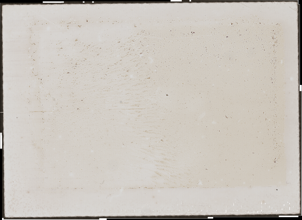
Unfortunately, because it is an entire slide containing cells, it is not possible to visualize in this mosaic image that is composed of about 25 to 33 thousand fields captured by the microscope. Each field has 1600×1200 px where each px has about 0.11×0.11 micrometers, the area of a cell nucleus has on average about 4610 pixels. For the construction of the dataset we selected fields that have at least one nucleus, below there is a field for better understanding.![]()
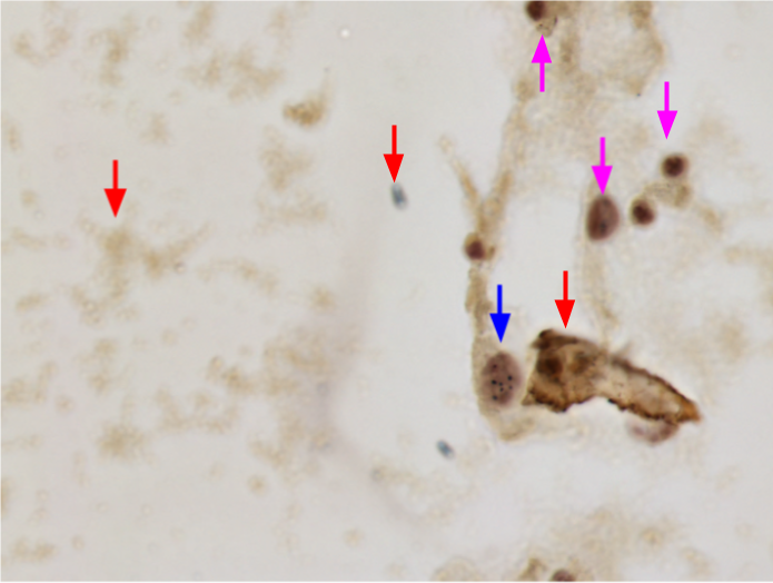
In this example of fields, there are parts indicated for a better explanation: the arrows in red indicate what is normally considered as the background of the image or dirt, this is considered noise, i.e. an area that is not of interest. The blue arrow indicates a nucleus that can be used and the pink arrows show out-of-focus nuclei that cannot be used because NORs within it cannot be identified. The following image highlights the NORs for better understanding.
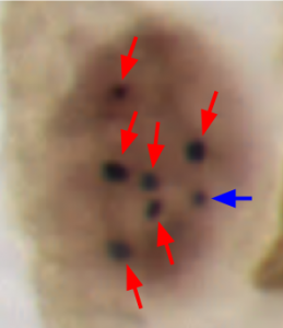
In this image highlighting a single nucleus is shown using the arrows, a cell considered healthy can have up to three NORs. These NORs are classified into two types, the only one highlighted by the blue arrow is called a satellite, a satellite as in the image is a single small and singular point. The NORs highlighted by the red arrows are called clusters they are larger and usually have granularity in their texture. Satellites have an average area of 100 pixels and clusters an area between 135 and 200 pixels.
Contents
Where was the data obtained from?
The images were obtained from examinations that were performed on women who were treated at the Gynecology and Colposcopy Outpatient Clinic of the University Hospital Professor Polydoro Ernani de São Thiago of Federal University of Santa Catarina (HU-UFSC). These women, presenting cytological alterations in the oncotic cytology of previous exams, were referred to the Ane Francyne Costa for a gynecological exam, colposcopy, and biopsy. This research was approved by the UFSC Research Ethics Committee (CEPSH), protocol number 57423616.3.0000.0121. All participating patients were previously approached and informed about the study objectives. Those who agreed to participate signed an Informed Consent Form.
Versions
- The dataset is available for download at https://arquivos.ufsc.br/d/373be2177a33426a9e6c/.
- This Version was published in CBMS 2020. Paper title: A Novel Approach on Segmentation of AgNOR-Stained Cytology Images Using DeepLearning
- It can be quoted using this Doi: 10.1109/CBMS49503.2020.00110
- The code and results can be accessed in this repository.
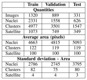
- Contains 2540 images (1600×1200 where each pixel is 0.111μm×0.111μm) from three different slides, having at least one nucleus per image.
- Convert the ground truth (the labels) to coco format
- Addition of the file
CCAgT_dataset_cocoFormat.jsoncontaining the dataset labels in coco format.
- In development
More about each version of the dataset
About the versions 1.x.x
I will be maintaining (still under construction) a public repository with codes (after publications) involving this dataset. A page of this repository can be accessed too.
Citing CCAgT
![]()
@inproceedings{AtkinsonAmorim2020,
doi = {10.1109/cbms49503.2020.00110},
url = {https://doi.org/10.1109/cbms49503.2020.00110},
year = {2020},
month = jul,
publisher = {IEEE},
author = {Joao Gustavo Atkinson Amorim and Luiz Antonio Buschetto Macarini and Andre Victoria Matias and Allan Cerentini and Fabiana Botelho De Miranda Onofre and Alexandre Sherlley Casimiro Onofre and Aldo Von Wangenheim},
title = {A Novel Approach on Segmentation of AgNOR-Stained Cytology Images Using Deep Learning},
booktitle = {2020 IEEE 33rd International Symposium on Computer-Based Medical Systems (CBMS)}
}